
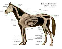
Understanding the anatomy of a horse’s skeleton is crucial for horse owners, veterinarians, and equitation enthusiasts alike. In this comprehensive article, we will delve into the intricate structure of the horse’s skeleton, comprising bones, teeth, vertebrae, ribs, and limbs.
The horse’s skeleton is composed of over 200 bones and can be divided into three main categories: head, neck, and body with limbs. Each anatomical region has unique characteristics and functions. For instance, the head contains the skull and teeth, while the neck supports the head and facilitates movement. The trunk includes the vertebral column, ribs, and pelvis, whereas the limbs are made up of bones such as the femur, tibia, and metatarsus.
A thorough understanding of the horse’s skeleton is essential for optimal performance, health, and overall well-being. Let us explore the intricate structure and functions of the horse’s skeleton in depth.
The Horse Skeleton: Structure and Functions
The horse’s skeleton is a robust and dense structure that enables the animal to withstand immense pressure and stress derived from its size and weight. This section will examine the anatomy of the horse’s skeleton, subdividing it into four main sections: skull and mandible, vertebral column, ribs and sternum, and limb bones.
Skull and Mandible
The horse’s skull is composed of numerous bones that fuse together to form a complex structure. The skull is robust and resilient, providing protection for the brain and sensory organs. The mandible, or lower jawbone, consists of two bones that unite at the base of the skull. The mandible is incredibly strong and capable of crushing tough and fibrous plant material.
The horse’s skull also features a unique arrangement of facial bones, including the maxilla, zygoma, and nasal bone. These bones form the structure of the face and are critical for maintaining eye alignment, breathing, and feeding.
Vertebral Column
The horse’s vertebral column comprises numerous vertebrae that extend from the base of the skull to the tail. The vertebral column is highly flexible, allowing for fluid movement and natural locomotion. The vertebrae can be divided into three main sections: cervical, thoracic, and lumbar.
Cervical Vertebrae
The cervical vertebrae are located in the neck region and consist of seven vertebrae. These vertebrae form a flexible joint that allows for subtle movements, enabling the horse to adjust its head position and facilitate feeding and drinking.
Thoracic Vertebrae
The thoracic vertebrae are situated in the upper back and comprise 13 vertebrae. These vertebrae form the main structural support of the trunk, allowing for upright posture and locomotion.
Lumbar Vertebrae
The lumbar vertebrae are located in the lower back and consist of five vertebrae. These vertebrae provide additional support to the vertebral column, enabling the horse to maintain balance and stability during movement.
Ribs and Sternum
The horse’s ribs are long and slender, extending from the vertebral column to the sternum. Ribs are connected to the vertebral column via articular connections. The sternum, or breastbone, consists of a series of flat bones located in the front portion of the thorax. The sternum plays a vital role in respiration, as it is connected to respiratory muscles.
The horse’s ribcage also features two pairs of ribs that attach to each other via costal cartilages. This unique arrangement provides additional support and flexibility to the vertebral column.
Limb Bones
The horse’s limbs comprise numerous bones that extend from the shoulder to the hoof. Limb bones can be divided into two main sections: upper limb and forelimb. The upper limb consists of one long bone called the humerus, while the forelimb comprises two long bones called radius and ulna.
Hooves are made up of multiple bones, including those in the carpus, metacarpus, and phalanges. The horse’s hoof is a unique structure composed of keratin-based tissue that provides shock absorption and traction during movement.
Understanding the structure and functions of the horse’s skeleton is crucial for optimal performance, health, and overall well-being.
Evolution and History
Horses have undergone an extraordinary evolutionary journey throughout millennia. The earliest known ancestor of the modern horse, Eohippus or Hyracotherium, lived around 55 million years ago during the Eocene epoch. This ancient species was only about 25 cm tall and had four toes on each front leg and three on the back legs.
Over time, horses have undergone numerous transformations that have led to the emergence of larger, more efficient species with fewer toes. Adaptations such as increased leg length and reduced toe number enabled early horses to become faster and more agile predators.
Adaptations Evolutionary
Horses’ evolution has been characterized by a range of adaptations that enable them to thrive in increasingly hostile environments. One key adaptation is the elongation of their legs, which allows for faster running and increased distance covered. Additionally, horses have developed larger teeth, better suited for grinding tough and fibrous plant material.
Other important adaptations include the emergence of a single hoof on each leg, enabling smoother movement and reducing injury risk, and improvements in vision, allowing them to detect predators at greater distances.
Horses’ unique evolutionary history has enabled them to become incredibly fast and resilient animals, capable of adapting to diverse environments and habitats.
Role in Veterinary Studies and Research
The horse’s skeleton plays a pivotal role in veterinary studies and research. Due to its complex structure, it is possible to identify various diseases and conditions affecting the horse.
Diseases of the Skeleton
Horses can be affected by an array of skeletal disorders, ranging from mild to severe. Common issues include arthritis, sacroiliac joint disease, and hoof wall abscesses. Early diagnosis through advanced diagnostic techniques is essential for providing optimal care.
Diagnostic Techniques
Veterinarians use a range of diagnostic techniques to identify skeletal abnormalities in horses. Radiography, ultrasound, and MRI are commonly employed methods for visualizing the horse’s skeleton and detecting potential issues.
Conservation and Display
Preparation of Horse Skeletons
Preparing a horse skeleton for display requires meticulous attention to detail and care. The first step involves cleaning the bones, which must be thoroughly washed and disinfected to remove any residual organic matter.
Once cleaned, the bones are dried and conserved in a dry, dust-free environment to prevent mold and fungi growth.
Mounting of Skeletons
After thorough preparation, it is possible to mount the skeleton. This operation requires specialized expertise and precision, as each bone must be positioned correctly to ensure stability and accuracy.
Displaying Horse Skeletons
Once mounted, horse skeletons can be displayed in museums or private collections. To ensure long-term conservation, it is crucial that the environment surrounding the display be carefully controlled in terms of temperature, humidity, and lighting.
Furthermore, it is essential to protect the skeleton from external factors such as dust or insects, which could cause damage.
In conclusion, the preparation, mounting, and display of horse skeletons demand specialized expertise and attention to detail. However, the end result can be a remarkable work of art that offers significant scientific and cultural value.
Additional Considerations
Understanding the Horse’s Skeleton: Clinical Applications
The study of the horse’s skeleton has numerous clinical applications in equine medicine. For instance, radiographic imaging can help diagnose fractures, joint disorders, and other skeletal abnormalities.
Additionally, studying the anatomy of the horse’s skeleton can inform surgical techniques and optimize treatment strategies for various conditions affecting the musculoskeletal system.
The importance of a thorough understanding of the horse’s skeleton cannot be overstated. By appreciating the intricate structure and functions of this remarkable animal, we can gain valuable insights into its behavior, physiology, and evolutionary history.
Conclusion
In conclusion, the horse’s skeleton is a complex and fascinating structure that plays a vital role in the overall health and well-being of these magnificent animals. By exploring the anatomy and functions of the horse’s skeleton in depth, we can appreciate the intricate mechanisms that enable horses to move, feed, and interact with their environment.
Whether for veterinary studies, conservation efforts, or equitation enthusiasts, understanding the horse’s skeleton is essential for providing optimal care, promoting welfare, and preserving the cultural significance of these incredible animals.


